The National Agriculture and Food Research Organization (NARO), in collaboration with Kazusa DNA Research Institute have successfully developed a high-throughput process flow to realize the three-dimensional (3-D) visualization of crop root system architecture (RSA) in soil through a simple, fast and non-destructive method using X-ray computed tomography (CT). RSA of a plant has a close relationship to its efficiency to absorb water and nutrients in soil and is therefore an important factor to influence the crop production. Nevertheless, diagnosing RSA of plants had been extremely labor-consuming, thus crop breeding to improve RSA had been practiced hardly before. By optimizing the conditions for the CT scanning as well as the image processing, we successfully visualized RSA of potted rice plant in less than fifteen minutes. This methodology is expected to be utilized widely in the agricultural field, from crop breeding to improve RSA, to the selection of the suitable variety for an individual farm soil through the root growth diagnoses.
Overview
Roots are essential organs for plants to grow and develop, thus they are significantly important in crop production. However, improvement of RSA had rarely become the purpose of breeding in crops such as rice and soybean when their roots are inedible, compared to the case in root vegetables. Although breeding usually requires examining several thousands to several tens of thousands of plants before selecting promising individuals, to dig up and to wash off the roots one by one is an overwhelmingly labor-consuming process that it is impossible to perform on large population size.
NARO and Kazusa DNA Research Institute joint-research team had applied X-ray CT which is normally used for the medical diagnoses and the non-destructive measurement of machine parts to develop a high-throughput flow for the 3-D visualization of potted rice RSA which requires only 12 minutes for the whole process: ten minutes for the CT scanning and reconstruction, and 2 minutes for the image processing. There had been past attempts to use X-ray CT for the RSA visualization, but they were excessively time-consuming, requiring as much as several hours and was not suitable method for mass examination.
The development of this analysis flow realized simple, fast and non-destructive 3-D visualization of RSA, which opened the door to breeding of rice, soybean and other crop plants for the improvement of RSA. Furthermore, it is expected to be applied widely to the agricultural field, to realize RSA visualization of crops beside rice, root growth diagnosing and so forth. The research paper was published online on May 11, 2020 in "Plant Methods", an international scientific journal.
Publication
Shota Teramoto, Satoko Takayasu, Yuka Kitomi, Yumiko Arai-Sanoh, Takanari Tanabata and Yusaku Uga. High-throughput three-dimensional visualization of root system architecture of rice using X-ray computed tomography. Plant Methods. 2020; 66 URL:https://doi.org/10.1186/s13007-020-00612-6
For Inquiries
Contact:http://www.naro.affrc.go.jp/english/inquiry/index.html
Reference Information
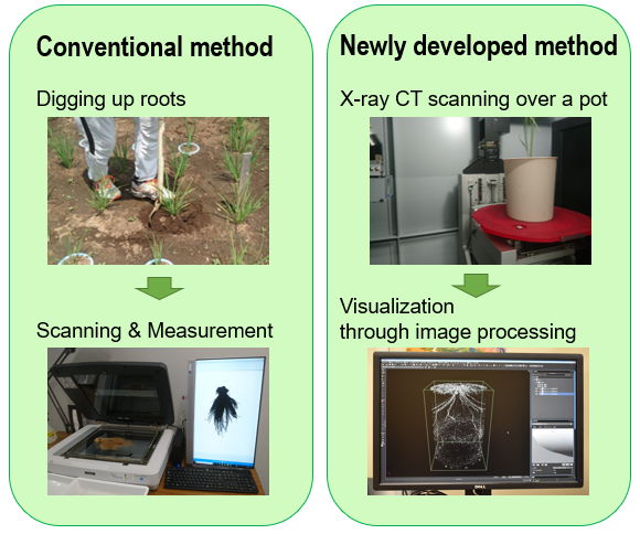
Fig.1 Comparison between the conventional method and the new method
The conventional method requires a process for roots to be dug up from the soil and washed off before the scanning and the measurement. The new method scans cultivated plant in a pot by X-ray CT directly and visualizes the RSA through fully automatic image processing. The new method requires less labor than the conventional method. Since it is non-destructive, sequent visualization to observe the development of RSA is even possible.
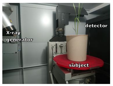
Fig.2 How X-ray CT scanning is performed
X-rays are projected from the generator, going through the subject on the turntable and striking the detector for the signals to be converted into an image. Scanning the subject from various angles enables the 3-D visualization inside the subject.
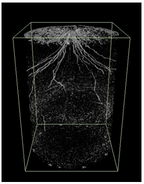
Fig.3 Visualization of rice RSA in the soil using X-ray CT
Visualization of rice RSA in soil using the newly developed method using X-ray CT. Roots are expressed as the linear white glow in the dark.
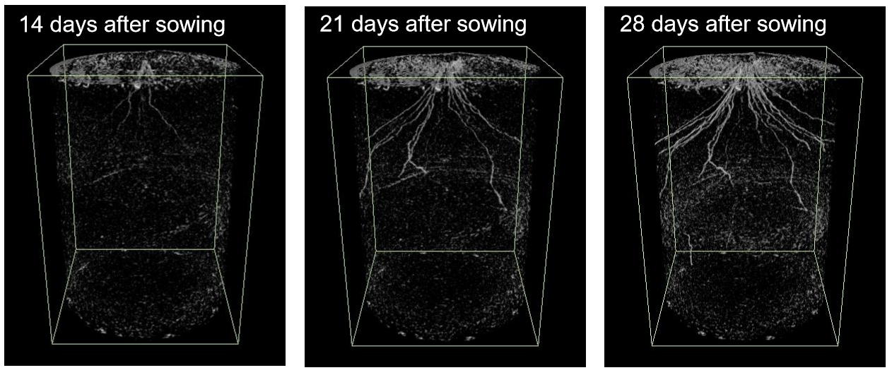
Fig.4 Chronological visualization of rice RSA development using X-ray CT
Three images are the rice RSA in different stages visualized by using the newly developed method. Root segments are too thin and hardly visible in the image on the left (14 days after sowing), but they gradually grow thicker as well as the whole structure grows as seen in image in the center (21 days after sowing), and even more so in image on the right (28 days after sowing).