The National Agriculture and Food Research Organization (NARO) had demonstrated that viewing a flower image affects the brain activity and thereby induce psychological and physiological reaction which supports recovery from psychological stress. The experiment put the subject group under a visual stress followed by showing a typical flower image which led to downregulation of negative emotions and decrease in elevated blood pressure and cortisol levels. Through this research, the mechanism of viewing flowers to reduce human stress was explained scientifically using psychology, physiology and neuroimaging techniques.
Overview
NARO, in collaboration with the University of Tsukuba, had clarified the mechanism of passive viewing of a flower image promoting recovery from stress by collecting evidence using psychological, physiological and neuroimaging methods.
The subject group was put under visual stress which proceeded by showing them a typical flower image leading to reduce their negative emotion as fear and disgust, and even shifted to a positive tone. Other physiological reactions were the decline of the elevated blood pressure by 3.4%, rather significant rate compared to viewing other images, and also the fall of the elevated cortisol level by 21 %.
Through the careful analysis of the brain activity, the team also showed that viewing the flower image induced the reduction of the amygdala activity relevant to emotion regulation. The subjects' attention on visual stressor had been automatically distracted by the sight of the flower image, causing the reduction of amygdala activity and decrease of elevated blood pressure and cortisol level. Our mission continues to investigate the effect of viewing fresh flowers on increasing health.
Publication
Viewing a flower image provides automatic recovery effects after psychological stress. Mochizuki-Kawai H, Matsuda I, Mochizuki S. (2020). Journal of Environmental Psychology.
https://doi.org/10.1016/j.jenvp.2020.101445
For Inquiries
Contact:http://www.naro.affrc.go.jp/english/inquiry/index.html
Reference Information
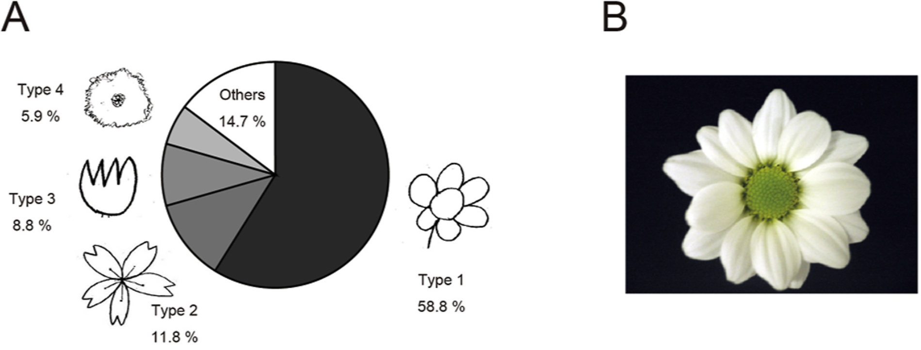
Fig. 1 Floral stimulus.
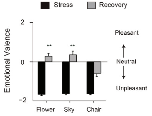
Fig. 2 Self-reported emotional valence. Mean rating scores of emotional valence during the presentation of a negative IAPS image (black bar) or after the presentation of one of three stimuli: flower, sky, or chair (gray bar). The changes in emotional valence were significantly larger in the flower and sky conditions than in the chair condition (P < 0.01). **P < 0.01.
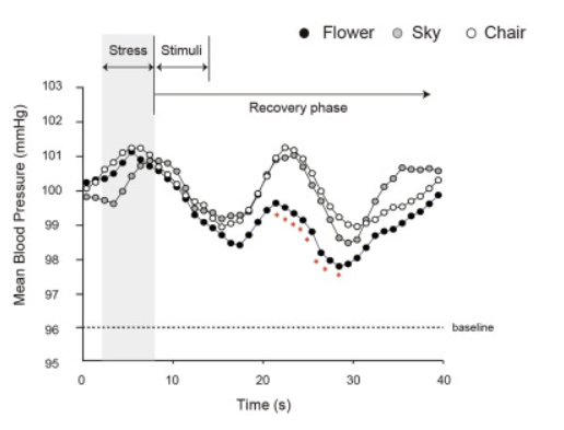
Fig.3 Changes in mean blood pressure (MBP) during Studies. The effect of viewing a negative IAPS image and subsequently viewing a flower image (solid circles), a sky image (gray circles), or a chair image (open circles) on MBP. The MBP during 8 s of the recovery phase was significantly lower in the flower condition than in the sky or chair condition (P < 0.05, marked by asterisks).
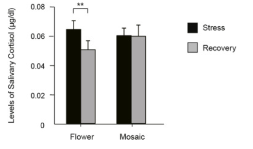
Fig.4 Changes in mean salivary cortisol levels.
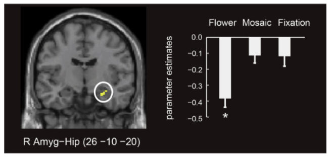
Fig. 5 Brain regions showing decreased activation during automatic emotional regulation induced by a flower image. Left: Right amygdala-hippocampus. Right: The decrease in the activation (mean parameter estimates) in the right amygdala-hippocampus was larger in the flower condition than in the mosaic and fixation conditions (P < 0.05). *P < 0.05.


